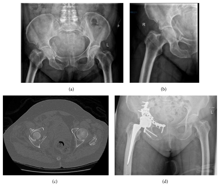
Figure 1.
Pre- and post-op images of a typical case involving fractures of both columns. (a, b) Initial AP pelvis and film of right hip. (c) Axial CT scan of the fracture showing displacement of both columns. (d) Postoperative X-ray. Both columns have been reduced and plated, and a hip replacement was performed.

