Figure 3.

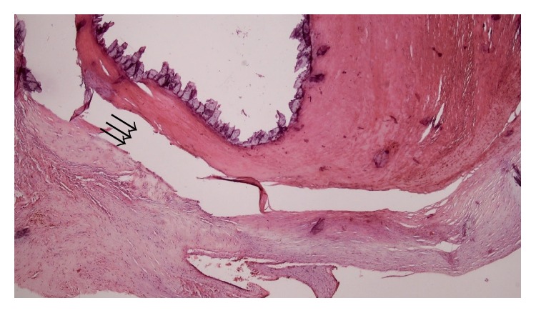
Histological examination showed dense calcification (shredded due to no decalcification) (arrows) in a background of amorphous degenerating fibrinous material (hematoxylin and eosin stain; ×40).

Histological examination showed dense calcification (shredded due to no decalcification) (arrows) in a background of amorphous degenerating fibrinous material (hematoxylin and eosin stain; ×40).