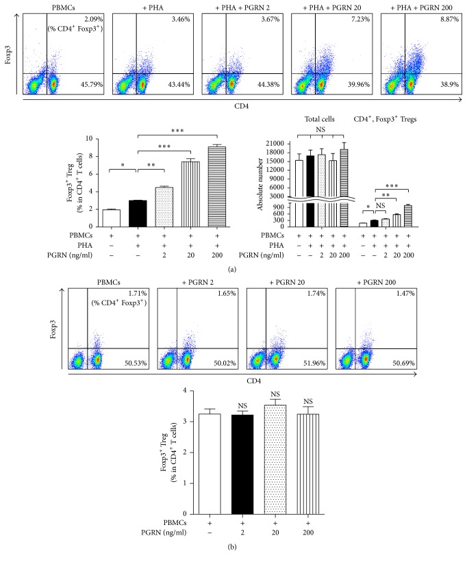
Figure 2.
PGRN enhances numbers of CD4+Foxp3+ Tregs under conditions of stimulation. hPBMCs were incubated with PHA plus PGRN at concentrations of 2, 20, and 200 ng/ml for 5 days (a). hPBMCs were cocultured with PGRN at concentrations of 2, 20, and 200 ng/ml for 5 days (b). Cells were collected and stained as described in Section 2. Stained cells were analyzed by flow cytometry. In (a), left bar graphs show the percentage of CD4+Foxp3+ iTreg in CD4+ T lymphocytes. Right bar graphs indicate cell numbers of total cells and CD4+Foxp3+ iTreg, respectively. Data are presented as the means ± standard deviations of three experiments performed in triplicate. ∗p < 0.05, ∗∗p < 0.01, ∗∗∗p < 0.001, or ns (not significant). Similar results were obtained in five independent experiments.

