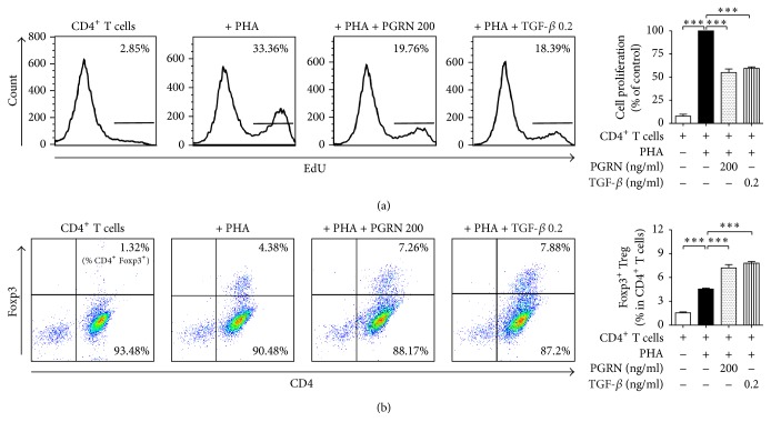
Figure 5.
TGF-β inhibits CD4+ T lymphocyte proliferation and induces iTreg formation as well as PGRN. CD4+ T lymphocytes were incubated with PHA in the presence of 200 ng/ml of PGRN or 0.2 ng/ml of TGF-β for 5 days. For the proliferation assay, cells that had incorporated EdU were counted by flow cytometry and Foxp3+ T lymphocytes among the CD4+ T lymphocytes were analyzed by flow cytometry. (a) shows the percentage of CD4+ T lymphocytes incorporated with EdU. (b) shows the percentage of CD4+Foxp3+ iTreg. Data are the means ± standard deviations of three experiments performed in triplicate. ∗∗∗p < 0.001. Similar results were obtained in four independent experiments.

