Figure 1.
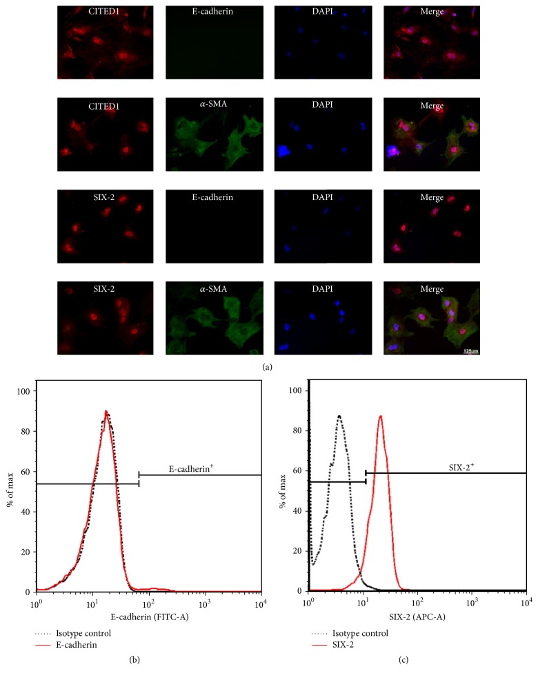
The isolated cells were identified as MMSCs by immunofluorescence double staining and flow cytometry. (a) The isolated cells were plated on chamber slide system and cultures in 21% O2 for 24 h, and then the cells were stained by immunofluorescence. The isolated cells coexpressed SIX-2 or CITED1 (markers of metanephric stem cell) and α-SMA (marker of mesenchymal cells). Both SIX-2 and CITED1 were located in the nucleus. α-SMA was expressed in cytoplasm. No E-cadherin (marker of epithelium) positive cells were observed. Bar is equal to 125 μm. (b)-(c) As the flow cytometry results shown, the percentage of E-cadherin cells is less than 1% and that of SIX-2 positive cells is over 90%. These results confirmed that the isolated cells were MMSCs.

