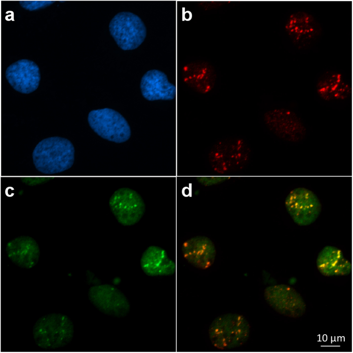
Figure 3. Induction GFP-RNF8 and γH2AX IRIF in cells irradiated with random α-particles.
(a) Hoechst33342 staining reveals the nuclear chromatin. (b) IRIF are visualized with γH2AX immuno-detection in fixed cells exposed to 239Pu source. (c) GFP-RNF8 is re-localized to the DNA damaged areas, and (d) the merged image shows the overlap of GFP-RNF8 and γH2AX 30 minutes after irradiation.

