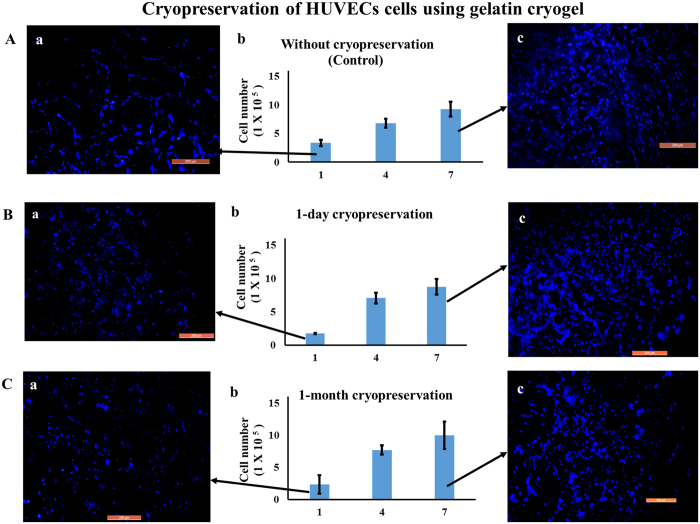
Figure 7. Cell proliferation and fluorescent microscopic images of HUVECs cells seeded on gelatin cryogel.
Post thawing cells were cultured in a CO2 incubator for 7 days (37 °C, 5% CO2). Cell proliferation assay of cells seeded gelatin cryogel (Ab) without cryopreservation (control), (Bb) 1 day and (Cb) 1 month cryopreservation. Nuclei staining at day 1 (Aa,Ba,Ca) and at day 7 (Ac,Bc,Cc) in case of control, after 1-day, and 1-month cryopreservation, respectively. Nuclei were stained with DAPI (blue). Scale bar: 200 μm; magnification: 100×.

