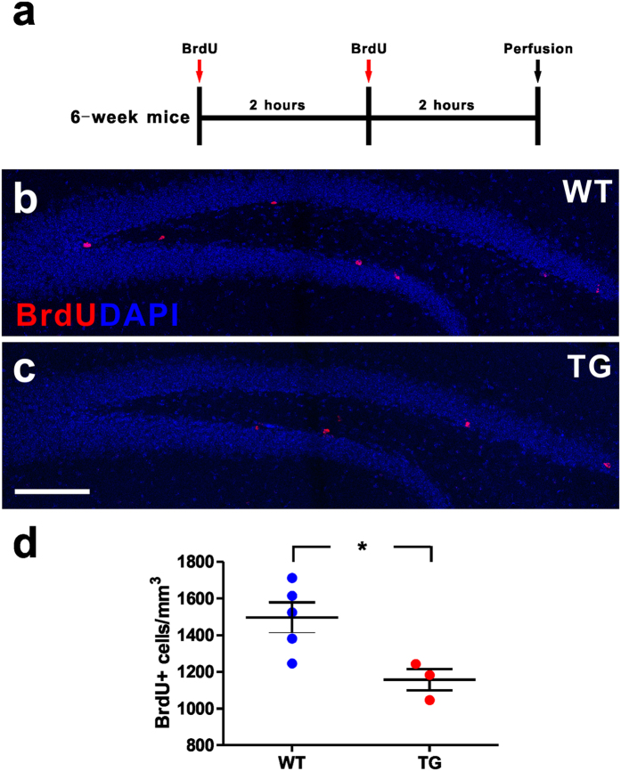
Figure 1. Reduced adult hippocampal dividing cells in MECP2 transgenic mice.
(a) Experimental scheme for assessing new dividing cells in the adult hippocampus. (b,c) Confocal microscopy images of the adult hippocampus showing dividing cells in the subgranular zone (SGZ) of wild type (WT) and MECP2 transgenic (TG) mice, displaying BrdU staining (red). The nuclear label DAPI is shown in blue. Scale bar: 200 μm. (d) Quantitative analysis of BrdU label density in granule cell layer. Values are Mean ± S.E.M (n = 5 WT; n = 3 TG; *P < 0.05, student’s t-test).

