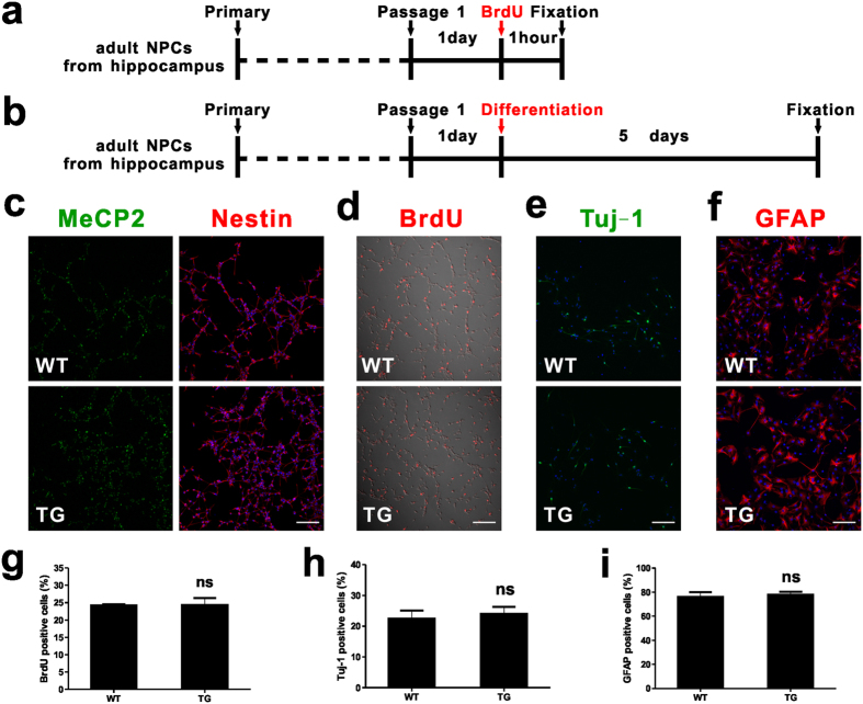
Figure 5. Proliferation and differentiation of adult hippocampal NPCs in vitro.
(a) Experimental scheme for assessing proliferation of NPCs in vitro. (b) Experimental scheme for assessing differentiation of NPCs in vitro. (c) Adult hippocampal NPCs culture under proliferating conditions expressed the neural progenitor cell marker Nestin (cytoplasmic, red); MeCP2 in green; Dapi in blue. Scale bar: 100 μm. (d) Both WT and TG NSCs incorporate the thymidine analog, BrdU, under proliferating conditions (BrdU, red). Scale bar: 100 μm. (e) Differentiation of adult NSCs into neurons (Tuj-1, green) by 1 μM RA and 5 μM forskolin. Scale bar: 100 μm. (f) Differentiation of adult NSCs into astrocytes (GFAP, red) by 1% FBS. Scale bar: 100 μm. (g–i) Quantitative analysis showing the percentages of BrdU+, Tuj-1+, and GFAP+ cells in WT and TG individuals respectively. Values are Mean ± S.E.M (ns: non-significant, student’s t-test).

