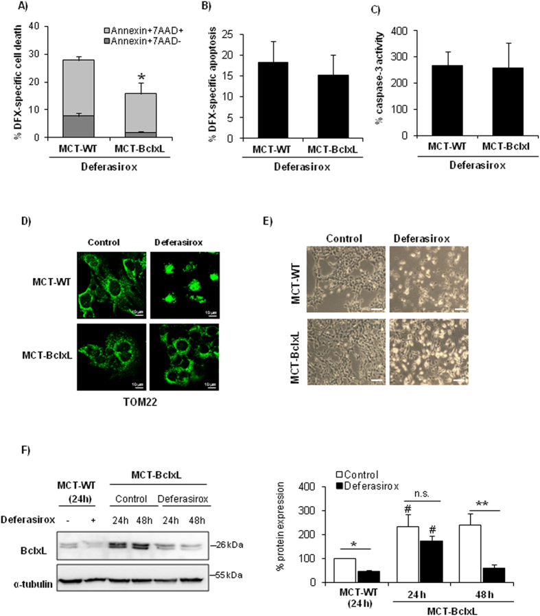
Figure 6. BclxL downregulation is involved in deferasirox-induced cell death.
Deferasirox toxicity was explored in BclxL-overexpressing MCT tubular cells (MCT-BclxL) and wild type cells (WT-MCT). (A) BclxL-overexpressing cells were protected from cell death induced by exposure to 10 μM deferasirox for 24 h, as assessed by flow cytometry of annexin V/7-AAD stained cells. Mean ± SEM of four independent experiments. *p < 0.03 vs control WT cells. (B) BclxL overexpression did not significantly decrease the presence of hypodiploid cells. Mean ± SEM of three independent experiments. (C) BclxL overexpression did not prevent caspase-3 activation induced by deferasirox. Mean ± SEM of three independent experiments. (D) TOM22 staining. MCT-BclxL cells, but not WT cells, preserve mitochondrial integrity in presence of deferasirox. Representative images of three independents experiments. Magnification x63. (E) Contrast phase microscopy photographs of MCT-WT and MCT-BclxL (original magnification x200, scale bars 200 μm). An increased number of cells that remain attached to the plate is observed in MCT-BclxL cells exposed to deferasirox. Representative images of three experiments. (F) BclxL protein expression in MCT-BclxL cells during exposure to deferasirox assessed by western blot. Mean ± SEM of three independent experiments. *p < 0.05 vs control MCT-WT; **p < 0.005 vs control MCT-BclxL; #p < 0.004 vs MCT-WT. Full-length blot is presented in Supplementary Figure 3.

