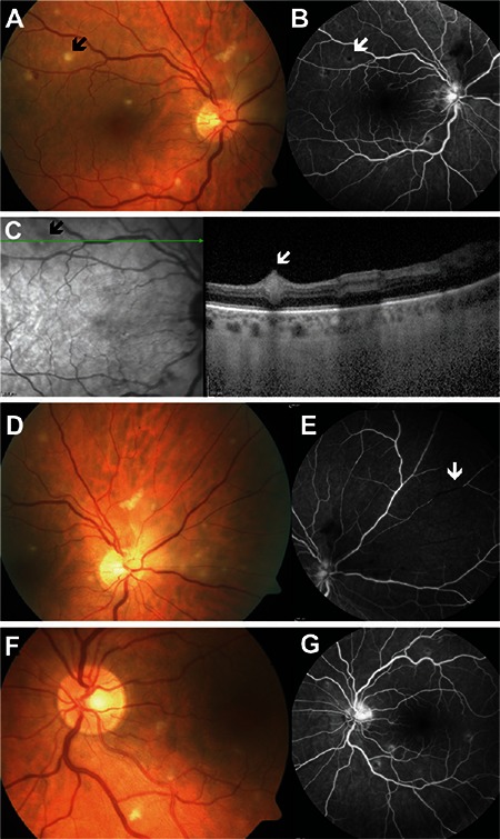
Figure 1. Imaging of patient 4 performed at time of presentation: right eye color fundus photographs (A and D), right eye fluorescein angiography (B and E), optical coherence tomography cross-section including retinal infiltrates in the superotemporal quadrant of the right eye (C), color photography of left eye (F), and fluorescein angiography image (G). Color fundus photography of the right eye shows multiple retinal infiltrates in the posterior pole and superonasal quadrant, and a superonasal area of retinal edema adjacent to the optic disc (A and D). Fluorescein angiography of the right eye shows partial staining of the optic disc, posterior pole retinal infiltrates with central hypofluorescence surrounded by hyperfluorescence, an area of retinal ischemia adjacent to the optic disc and arteriole filling defect (arrow) in the superonasal quadrant (B and E). Optical coherence tomography corresponding to the retinal infiltrates in the superotemporal quadrant of the right eye (indicated by arrows in A, B and C) shows focal hyperreflective retinal thickening (C). Color fundus photography of the left eye revealed multiple retinal infiltrates at the posterior pole (F). Fluorescein angiography of the left eye shows partial staining of the optic disc and posterior pole retinal infiltrates with central hypofluorescence surrounded by hyperfluorescence (G).

