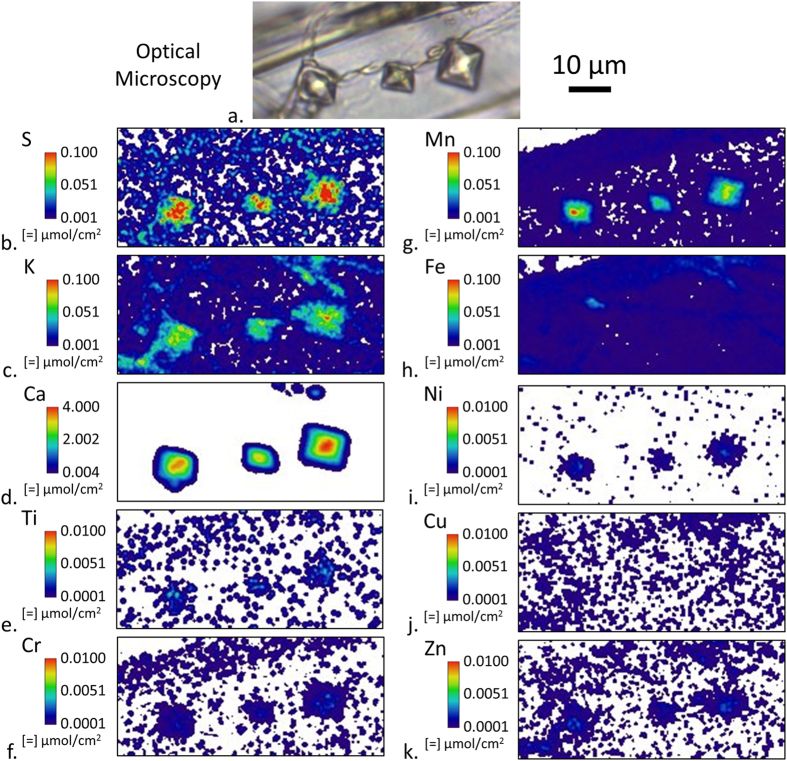
Figure 5.
Optical microscopy (a) and submicron resolution XFM ion maps of three oxalate crystals within a 2-μm-thick tangential-longitudinal section that was exposed to a Serpula lacrymans inoculated feeder strip in a soil block test for 20 days. Between elements, intensities were scaled to optimize visualization of ion distribution.

