
Fig. 2.
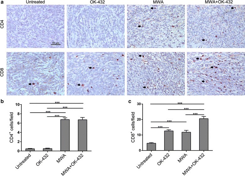
OK-432 increased infiltration of cytotoxic CD8+ T-cell into tumors after MWA. a representative microphotographs of CD4 and CD8 staining in each group. Immunohistochemical staining was performed on tumor specimens that were harvested 7 days after treatment. Original magnification, ×400. Bar, 50 μm. b, c Density of CD4+ and CD8+ cells was determined. Five random areas within a section were chosen and counted at 400-fold magnification. Columns, mean; error bar, SEM. ***P < 0.001. Data were pooled from two independent experiments with five mice per group