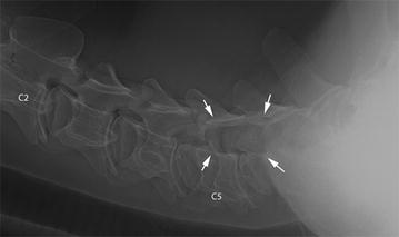
Fig. 1.

Laterolateral radiographic view of the cervical vertebral column. Laterolateral radiographic view of the cervical vertebral column of a 9.5-month-old Red Holstein heifer. The second (C2) and fifth (C5) vertebrae are labelled. A lytic lesion at the level of C5 and C6 (arrows) superimposed on the vertebral canal corresponds to the lateral parts of these vertebrae. The lesion has a thin sclerotic rim cranially
