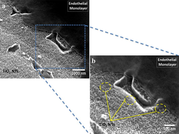
Fig. 9.

SEM micrographs highlighting the formation of endothelial monolayer on the NTs in a cross-section view after 24 h. a Endothelial layer (white dotted lines) growing on the NT surface at low magnification; and b Endothelial monolayer (white dotted lines) spread on the NTs (yellow circles) at higher magnification
