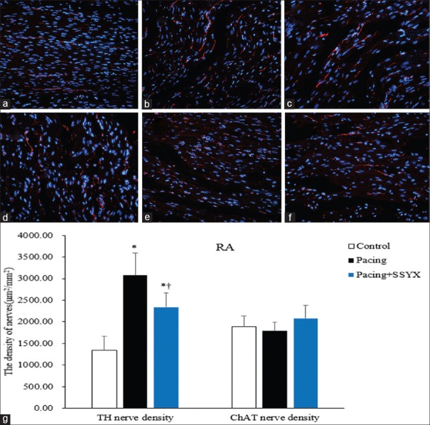
Figure 3.
Histological findings of TH and ChAT RA nerves in all groups. Representative images of TH immunofluorescence staining (original magnification, ×400) of RA in the control group (a), the pacing group (b), and the pacing + SSYX group (c). Exemplary pictures of ChAT immunofluorescence staining of RA in the control group (d), the pacing group (e), and the pacing + SSYX group (f) and statistical chart of TH and ChAT RA nerve (g). *P < 0.001 versus control group; †P < 0.05 versus pacing group. Blue: Nucleus of cardiomyocyte; Red: TH-positive and ChAT-positive nerves. TH: Tyrosine hydroxylase; ChAT: Choline acetyltransferase; RA: Right atrium; SSYX: Shensong Yangxin.

