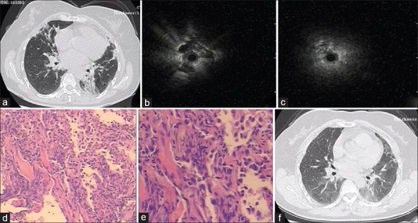
Figure 1.
Clinical and pathological images in a 46-year-old patient. (a) Computed tomography imaging showed bilateral multiple alveoli filling. (b) Radial probe endobronchial ultrasound showing the peripheral pulmonary lesion as a hypoechoic structure in the left lower and right middle lesion. (c) Radial probe endobronchial ultrasound showing the peripheral pulmonary lesion as a hypoechoic structure in the right middle lesion. (d) Organizing pneumonia identified in this patient (H and E, ×50). (e) Masson bodies apparent in the alveoli (H and E, ×100). (f) After 1-month follow-up, chest computed tomography scans showed that lesions in the right middle and left lower lobe were reduced.

