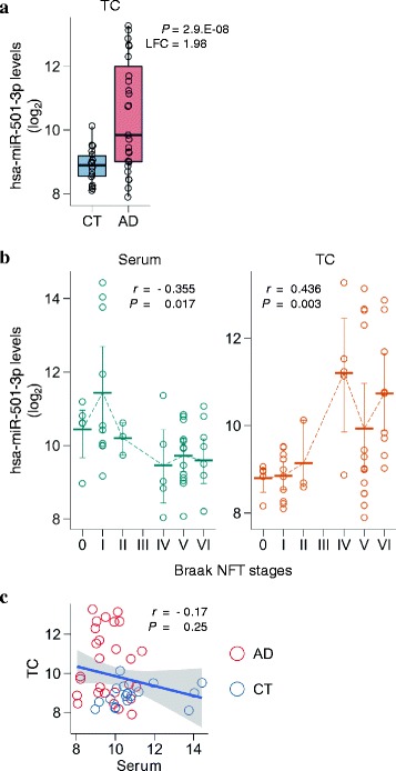
Fig. 2.

Deregulated hsa-miR-501-3p not only in the serum but also in the brain of the same individuals over the course of Alzheimer’s disease (AD) progression. a Brain hsa-miR-501-3p levels prominently upregulated in the AD brains of the ROW discovery set. The levels of hsa-miR-501-3p in the temporal cortex were analyzed using next-generation sequencing (NGS). Normalized read counts from the NGS data were converted to a log2 scale and plotted against the disease status. Box plots display the distributions of data in the same way as in Fig. 1a. CT: control; LFC: log2 fold change; P: p-value adjusted by the Benjamini–Hochberg procedure for multiple testing correction; TC: temporal cortex. b Significant correlation of either serum hsa-miR-501-3p levels or brain hsa-miR-501-3p levels with Braak NFT stages in the ROW discovery set. While serum hsa-miR-501-3p levels negatively correlated with Braak NFT stages (Spearman’s r = −0.355, P = 0.017), brain hsa-miR-501-3p levels positively correlated with Braak NFT stages (Spearman’s r = 0.436, P = 0.003). Normalized read counts from the NGS data were converted to a log2 scale and plotted against Braak NFT stages. Each thick bar shows the mean value of hsa-miR-501-3p levels in a Braak NFT stage. Each error bar shows a 95% confidence interval of hsa-miR-501-3p levels in a Braak NFT stage. TC: temporal cortex. c Negative correlation between serum hsa-miR-501-3p levels and brain hsa-miR-501-3p levels in the same individuals of the ROW discovery set. The levels of hsa-miR-501-3p between serum and brain of the same individuals were insignificantly yet negatively correlated (Spearman’s r = −0.17, P = 0.25). Normalized read counts of hsa-miR-501-3p levels were converted to a log2 scale and plotted. The x-axis and y-axis show serum hsa-miR-501-3p levels and brain hsa-miR-501-3p levels, respectively. The blue line shows a linear regression line, and a shaded gray area around the line represents 95% confidence intervals. Red circles and blue circles show the hsa-miR-501-3p levels of AD patients and control subjects, respectively. CT: control; TC: temporal cortex
