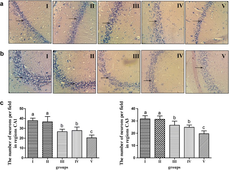
Fig. 2.

Morphologic changes of hippocampus stained by Nissl staining using a light microscope (magnification 400×) and counting the results. a Nissl staining of region CA1 of hippocampus in different groups. b Nissl staining of region CA3 of hippocampus in different groups. c The number of neurons per field in regions CA1 and CA3 of hippocampus. n=20, values are expressed as the mean ± SD. Bars without a common superscript letter differ significantly (P < 0.05)
