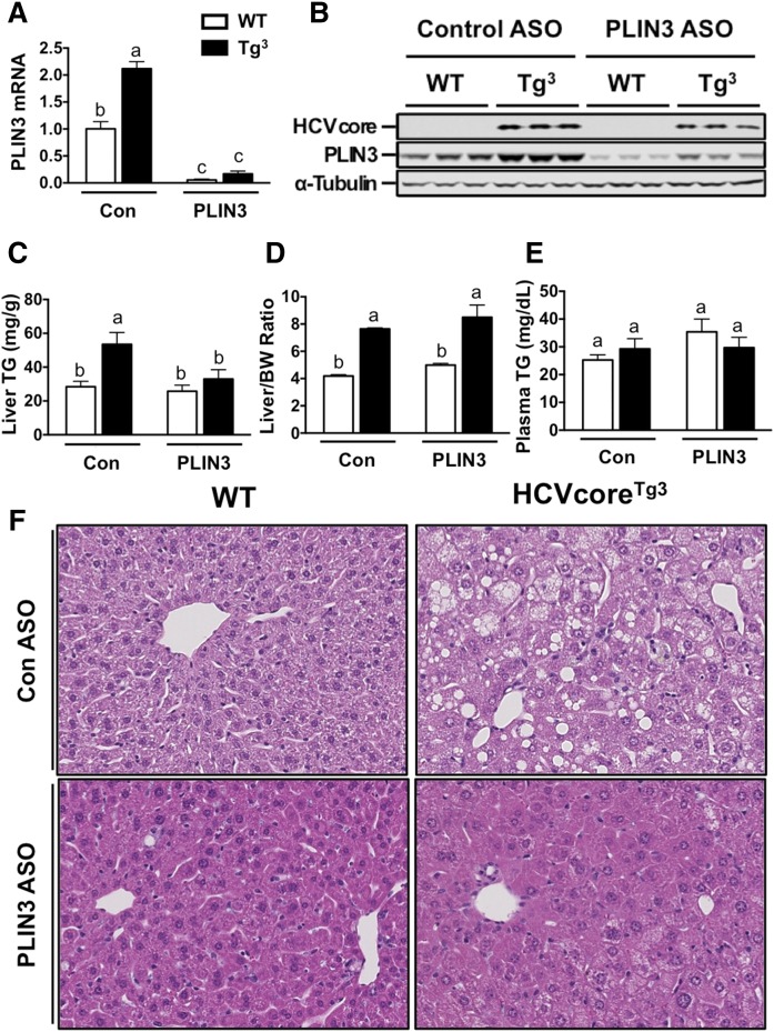
Fig. 4.
ASO-mediated knockdown of PLIN3 expression decreases steatosis in HCVcoreTg3 mice on chow diet. At 8 weeks of age, female WT and HCVcoreTg3 mice were treated with either control (Con) or PLIN3 ASO for 8 weeks while being maintained on a chow diet. A: Relative levels of liver PLIN3 mRNA were quantified by real-time PCR, normalized to levels of cyclophilin A, and expressed relative to levels in WT mice given control ASO (n = 4 per group). B: PLIN3 protein expression determined by Western blot analysis of liver homogenates. C: Enzymatic determination of TGs from liver lipid extract (n = 4–6 per group). D: Liver size (expressed as a ratio to body weight) of mice at necropsy. E: Measurement of plasma TGs determined enzymatically. F: Representative pictures of H&E staining performed on fixed liver sections (20× magnification). All hepatic lipid values were normalized to tissue weight. Data shown represent mean ± SEM from four to six mice per group. Levels not connected by the same letter are significantly different.

