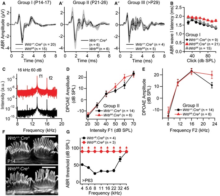
Figure EV3. Wrb disruption in hair cells causes a synaptopathic hearing impairment (related to Figs 3 and 7).

-
A–A″ABR wave I amplitudes of Wrb +/+:Cre A and Wrb fl/fl control animals of groups I‐III are normal in transgenic animals lacking Cre expression.
-
BABR latencies of group I animals are increased in Wrb fl/fl:Cre A when compared to Wrb +/+:Cre A and Wrb fl/fl control animals.
-
CRepresentative example frequency spectra of DPOAEs at 10.67 kHz measured from Wrb fl/fl:Cre A and Wrb +/+:Cre A mice to primary tones of 13.3 (70 dB) and 16 kHz (60 dB).
-
D, ENo statistically significant differences in (D) mean DPOAE amplitudes at increasing sound intensities in Wrb fl/fl:Cre A and Wrb +/+:Cre A (exemplary at 16 kHz) or (E) across the entire measured frequency range (at intensities of 70 dB (f1) and 60 dB (f2)) could be observed, indicating unaltered cochlear amplification and OHC function in the mutants.
-
FIHC hair bundles appear normal in Wrb fl/fl:Cre A animals as assessed with fluorophore‐conjugated phalloidin stainings at P14. Scale bar: 5 μm.
-
GLack of ABR in > P63 Wrb fl/fl:Cre B mice across the entire frequency range. Upwards pointing arrowheads indicate thresholds exceeding the maximum speaker output of 90 dB.
