-
A
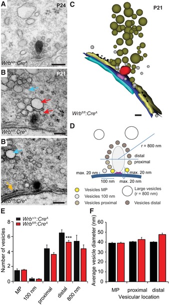
Representative electron micrograph of a Wrb
+/+:Cre
A IHC ribbon synapse. Scale bar: 120 nm.
-
B, B′
In Wrb
fl/fl:Cre
A (depicted are two consecutive ultrathin sections) IHCs, accumulations of large partially amorphous vesicles were observed close to ribbon synapses (red arrows), but also further away in the cytoplasm (cyan arrows). Moreover, cisternal structures resembling (r)ER are found unusually close to the ribbon (orange arrow in B′). Scale bar: 120 nm.
-
C
3D serial reconstruction of the ribbon synapse depicted in (B, B′, using “Reconstruct” software). Scale bar: 100 nm.
-
D
Schematic illustration of the quantitative analysis performed on random ultrathin sections, results shown in (E) and (F).
-
E
Quantification in random ultrathin sections (Wrb
+/+:Cre
A
n = 28, from two animals; Wrb
fl/fl:Cre
A
n = 44, from three animals) of the numbers of membrane‐proximal “MP” vesicles (in close proximity to membrane and ribbon); vesicles within a 100 nm range along the active zone membrane; vesicles at the lower “proximal” and the upper “distal” half of the ribbon as well as vesicles > 70 nm (“large vesicles”) in a radius of 800 nm around the ribbon. The vesicle number was significantly reduced in Wrb
fl/fl:Cre
A IHCs at the distal part of the ribbon, but not at the presynaptic membrane or proximal ribbon part. Data are represented as means ± SEM. Student's two‐sample t‐test, ***P < 0.001.
-
F
Average vesicle diameters of the membrane‐proximal vesicles and the vesicles at both halves of the ribbon. The mean vesicle diameter is unchanged in Wrb
fl/fl:Cre
A IHCs. Data are represented as means ± SEM.

