
Figure 1.
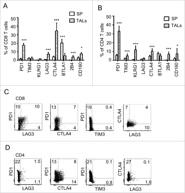
Multiple immune checkpoints are expressed on TALs of murine ovarian cancer microenvironment. Pooled data of eight inhibitory receptor expression on CD8+ (A) and CD4+ (B) TALs from tumor-bearing mice. IE9mp1 tumor cells (1×10⁷) were injected intraperitoneally. TALs were isolated at day 25 (Materials and Methods) and stained for surface expression of the checkpoints and analyzed with flow cytometry. Expression of eight checkpoints in T cells from splenocytes is shown for comparison. Data were obtained from 10 animals and are representative of two independent experiments. Data were analyzed using GraphPad Prism 6. Error bars represent SD. Statistical significance was determined by Student's t-test. *p < 0.05; **p < 0.01; ***p < 0.001. Examples of CD8+ (C) and CD4+ (D) TALs coexpressing two checkpoint molecules are shown. Representative flow cytometry analysis of TALs stained with Live/Dead dye, mAbs to CD4+, CD8+, PD-1, LAG-3, TIM-3, and CTLA-4. Dot plot analyses were gated on live cells, then on CD8+ or CD4+ and showed percentages of single and double stained PD-1, LAG-3, CTLA-4, and TIM-3-positive cells.