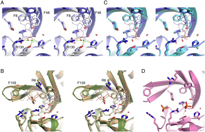
Fig 2. Crystal structures of LigT complexed with different active-site ligands.
A. Comparison between apo LigT (white) and the 2′-AMP complex (blue). Interactions of the ligand phosphate group are shown as orange dashed lines and those of the nucleophilic water molecule (green) in the apo structure as green dashed lines. B. Complexes with NADP+ (light brown) and ATP (green). Note the stacking of the NADP+ nicotinamide ring against Phe159 and the coordination of the 5′-phosphate in both ligands by Arg6. C. Binding modes of 2′-AMP (blue) and 3′-AMP (light blue). The same recognition elements are at play, but especially the ribose ring binds differently. D. Cocrystallization with tRNA resulted in a complex with two phosphate ions, one in the active site and the other one in a nearby pocket surrounded by Arg residues.

