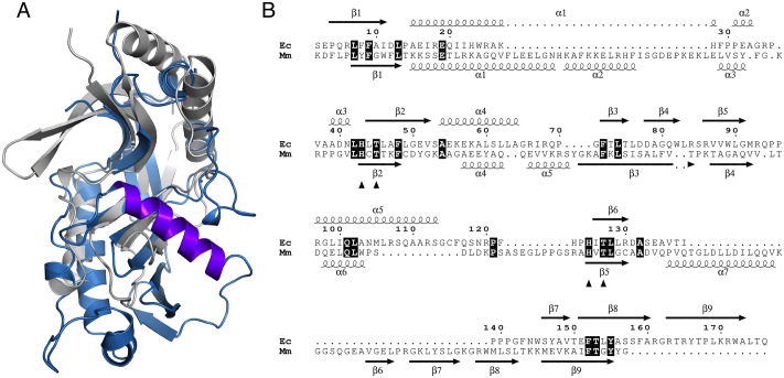
Fig 4. Comparison of CNPase and LigT.
A. Structural superposition of E. coli LigT (white) and mouse CNPase (blue). Specifically note the unique helix α7 (dark blue) in CNPase, lining the CNPase active site, and blocking access of nucleophiles larger than water. B. Structure-based sequence alignment of LigT (Ec) and CNPase (Mm).

