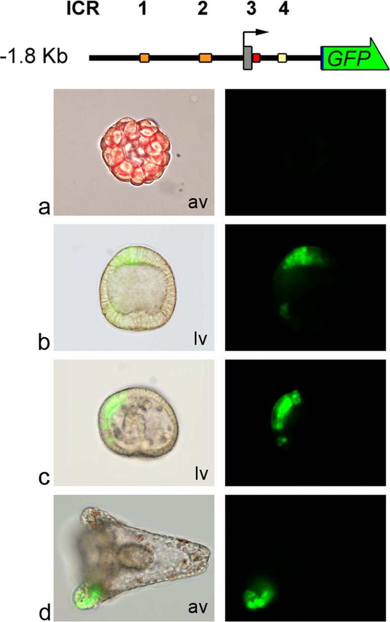
Fig 1. Expression of the -1.8KbGFP transgene construct during P. lividus embryo development.

At the top is a schematic structure (drawn to scale) of the Pl-Tuba1a -1.8KbGFP reporter construct. The bent arrow indicates the TSS. A grey box represents the first exon (5’UTR and ATG start codon). Downstream of the first exon there are the first intron and two codons of the second exon. Coloured boxes indicate the four ICRs. For sake of simplicity, only the section of ICR3 inside the intron is shown. The arrowed green box represents the GFP reporter gene cloned in frame with the alpha tubulin codons. Left: triple-merged images (bright-field, GFP fluorescence and Texas Red fluorescence-a) or merged fluorescence and bright-field images (b, c, d). Right: GFP fluorescence images from microinjected embryos. × 20 magnification. (a) 32-cell stage; (b) Blastula stage; (c) Gastrula stage; (d) Pluteus stage. Lv: lateral view; av: animal view.
