
Fig 4. Transgene loss of expression by intron deletion.
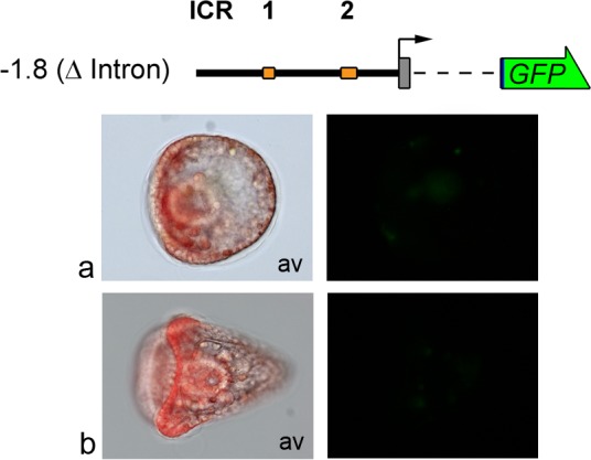
Embryos were microinjected with the -1.8(ΔIntron)GFP construct. Left: triple-merged images (bright-field, GFP fluorescence and Texas Red fluorescence) proving embryos are microinjected. Right: GFP fluorescence images. Structure and conventions are the same as in Fig 1. (a) Gastrula stage; (b) Pluteus stage. Av: animal view.