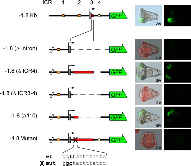
Fig 5. ICR3 and ICR4 function: spatial expression.
In these construct depictions, ICR3 was enlarged to show deletion details (not to scale). Left: schematic pictures of GFP reporter constructs. Structure and conventions are the same as in Fig 1. Right: merged fluorescence and bright-field images or triple-merged images and fluorescence images from microinjected pluteus stage embryos are shown to observe GFP localization. × 20 magnification. Av: animal view; ov: oral view.

