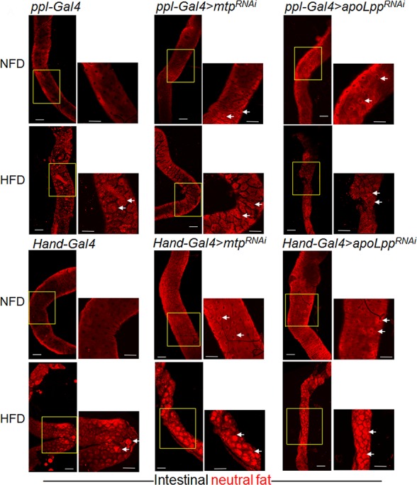
Fig 4. Inhibition of Mtp or apoLpp in fat body or cardiomyocytes promotes intestinal lipid accumulation on normal food diet and high fat diet.

(A-C’) Representative confocal images of Nile Red-stained lipid droplets in the unfixed intestines of third instar larvae on NFD. (A, A’), control larvae (ppl-Gal4); (B, B’), larvae with fat body-specific KD of mtp (ppl-Gal4>mtpRNAi); and (C, C’), larvae with fat body-specific KD of apoLpp (ppl-Gal4>apoLppRNAi). (A, B, C) Lower magnification (10X) images with scale bars representing 100 μm. (A’, B’, C’) Insets represent the magnified (20X) portion of the intestines (yellow boxes) with scale bar representing 70 μm. Arrows in insets indicate lipid droplets within the enterocytes. (D-F’) Representative confocal images of Nile Red-stained lipid droplets in the unfixed intestines of third instar larvae on HFD. (D, D’), control larvae (ppl-Gal4); (E, E’), larvae with fat body-specific KD of mtp (ppl-Gal4>mtpRNAi); and (F, F’), larvae with fat body-specific KD of apoLpp (ppl-Gal4>apoLppRNAi). (D, E, F) Lower magnification (10X) images with scale bars representing 100 μm. (D’, E’, F’) Insets represent the magnified (20X) portion of the intestines (yellow boxes) with scale bar representing 70 μm. Arrows in insets indicate lipid droplets within the enterocytes. (G-I’) Representative confocal images of Nile Red-stained lipid droplets in the unfixed intestines of third instar larvae on NFD. (G, G’), control larvae (Hand-Gal4); (H, H’), larvae with cardiomyocyte-specific KD of mtp (Hand-Gal4>mtpRNAi); and (I, I’), larvae with cardiomyocyte-specific KD of apoLpp (Hand-Gal4>apoLppRNAi). (G, H, I) Lower magnification (10X) images with scale bars representing 100 μm. (G’, H’, I’) Insets represent the magnified (20X) portion of the intestines (yellow boxes) with scale bar representing 70 μm. Arrows in insets indicate lipid droplets within the enterocytes. (J-L’) Representative confocal images of Nile Red-stained lipid droplets in the unfixed intestines of third instar larvae on HFD. (J, J’), control larvae (Hand-Gal4); (K, K’), larvae with cardiomyocyte-specific KD of mtp (Hand-Gal4>mtpRNAi); and (L, L’), larvae with cardiomyocyte-specific KD of apoLpp (Hand-Gal4>apoLppRNAi). (J, K, L) Lower magnification (10X) images with scale bars representing 100 μm. (J’, K’, L’) Insets represent the magnified (20X) portion of the intestines (yellow boxes) with scale bar representing 70 μm. Arrows in insets indicate lipid droplets within the enterocytes.
