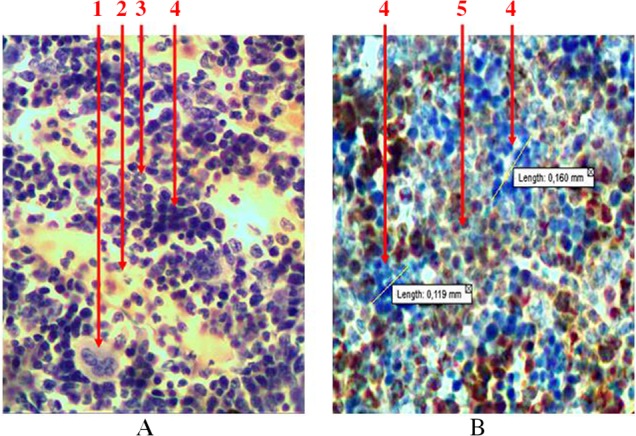
Fig. 6.

Longitudinal section of rat femur. 1 – megalokaryocyte, 2 – sinusoid, 3 – ring-shaped neutrophiles, 4 – erythroblastic insula, 5 – granulocytopoiesis area. (A) Hematoxylin and eosin staining and (B) immunohistochemical study of receptors for myeloperoxidase. Magnification 400×
