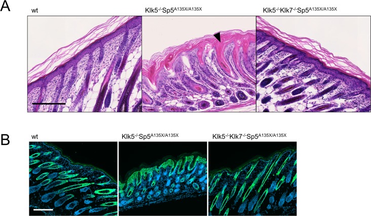
Fig 6. Histological analysis of epidermis structure at P5.
(A) Hematoxylin and eosin stained dorsal skin from wt, Klk5-/-Sp5A135X/A135X, and Klk5-/-Klk7-/-Sp5A135X/A135X mice at P5. Klk5-/-Sp5A135X/A135X pups show acanthosis and both the distribution and orientation of hair follicles were distorted. The follicles exhibit severe defects manifested by severe hyperkeratosis of isthmus and infundibulum (black arrowhead). The skin in Klk5-/-Klk7-/-Sp5A135X/A135X mice was comparable to wt mice. Scale bar, 200 μm (B) Skin sections from P5 pups were stained using anti-Keratin6 antibody. Klk5-/-Sp5A135X/A135X show hyperproliferation of keratinocytes, which is rescued in Klk5-/-Klk7-/-Sp5A135X/A135X. Scale bar, 200 μm.

