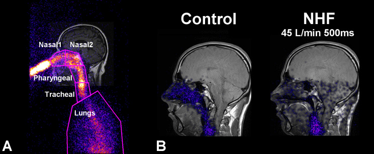
Fig. 1.
Lateral gamma camera image of nasal 81mKr gas inhalation overlaid on the coronal MRI image of a volunteer during breath holding. A: definition of anterior (Nasal1), posterior (Nasal2), pharyngeal, tracheal, and lung ROIs. B: visualization of 81mKr gas distribution 500 ms after the application of NHF at a rate of 45 l/min (right) compared with the control (left) shows fast clearance of the tracer gas in the upper airways. The control measurement without cannula flow shows stable 81mKr gas concentration.

