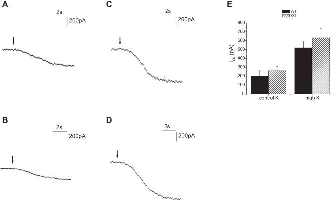
Fig. 12.
TPNQ-sensitive currents in Sgk1+/+ and Sgk1−/− mice. Whole cell currents were recorded at a membrane potential of 0 mV from principal cell of CCDs. TPNQ was added at the arrows. A: Sgk1+/+ mouse fed a control diet. B: Sgk1−/− mouse fed a control diet. C: Sgk1+/+ mouse fed a high-K diet D: Sgk1−/− mouse fed a high-K diet. E: quantitation of TPNQ-sensitive currents (ISK). Data are represented as means ± SE for 16–23 cells.

