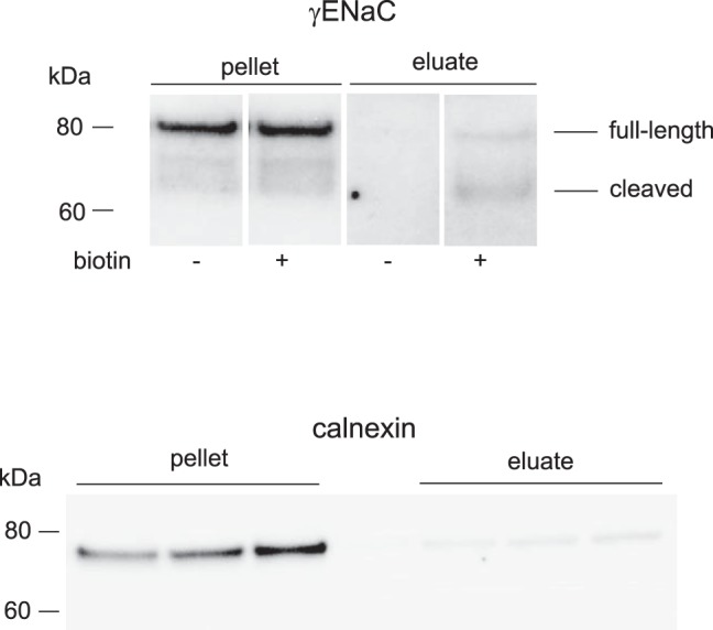
Fig. 6.

Biotinylation of mouse kidney surface proteins. Top: Western blot of kidney microsomes and neutravidin eluates from mouse kidneys perfused with or without biotin probed with antibodies against γENaC. Lanes were loaded with 50 μg of total microsomal protein, or eluates corresponding to 600 μg of total microsomal protein. Shown are 4 noncontiguous lanes of the same film of a single blot with uniform image processing. Intervening lanes are omitted for clarity. Bottom: Western blot of kidney microsomes and neutravidin eluates from mouse kidneys perfused with biotin probed with antibodies against calnexin. Lanes were loaded with 30 μg of total microsomal protein, or eluates corresponding to 600 μg of total microsomal protein.
