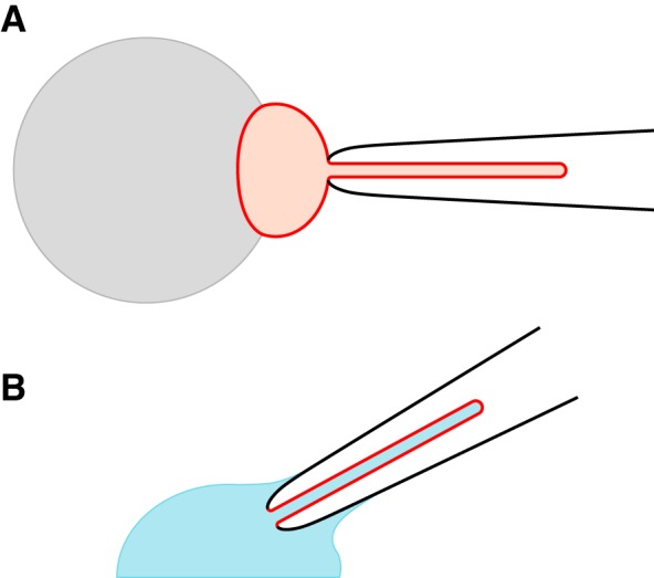
Fig. 1.

Configurations for recording ciliary transmembrane currents (30). A: recording from a cilium that remains attached to its cell. On a glass-coated bead (gray), renal epithelial cells are grown to confluence. Just one cell is shown (red). Suction from the recording pipette (black) pulls the cell’s primary cilium into the pipette. The pipette contains an external solution, as does the bath surrounding the cell. B: recording from a cilium detached from the cell. From the attached state (A), the pipette is lifted briefly into the air, which breaks off the cell body and leaves the excised cilium in the pipette. The pipette is quickly immersed in a cytoplasmic solution (blue), which diffuses into the cilium. In both configurations, currents seen principally reflect ciliary channels. The attached configuration (A) was used only for the work shown in Fig. 2; all other experiments used excised cilia (B). The drawings are not to scale.
