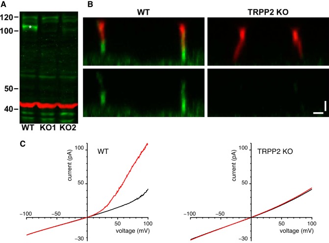
Fig. 9.
TRPP2 knockout (KO) mIMCD-3 cell lines. A: Western blot for TRPP2 protein with 40 µg of whole cell lysate from mIMCD‐3 wild-type (WT) and two TRPP2 KO cell lines. The band of TRPP2 protein (green, *) is present in the WT but not in the KO cells. β-Actin (red) was immunostained as a control for loading. Molecular mass markers are in kDa. B: immunostaining of mIMCD‐3 cells with the ciliary marker (37) anti-acetylated α-tubulin (red, top) and with anti‐TRPP2 (green, all images) shows that the TRPP2 KO cells have lost ciliary labeling for TRPP2. Each image is an XZ maximum intensity projection of a stack of XY images acquired at a range of Z depths on a confocal microscope. Bars, 1 μm. C: absence of current activated by 3 µM cytoplasmic Ca2+ in cells lacking TRPP2. For each cilium, macroscopic currents were determined by averaging currents from 10 to 40 voltage ramps. High-K+ external and cytoplasmic solutions were used; the cytoplasmic solutions contained 0.1 µM free Ca2+ (black) or 3 µM free Ca2+ (red). Results shown are averages from 9 cilia (wild-type with detectable large-conductance channels, at left) or 10 cilia (TRPP2 knockout, at right).

