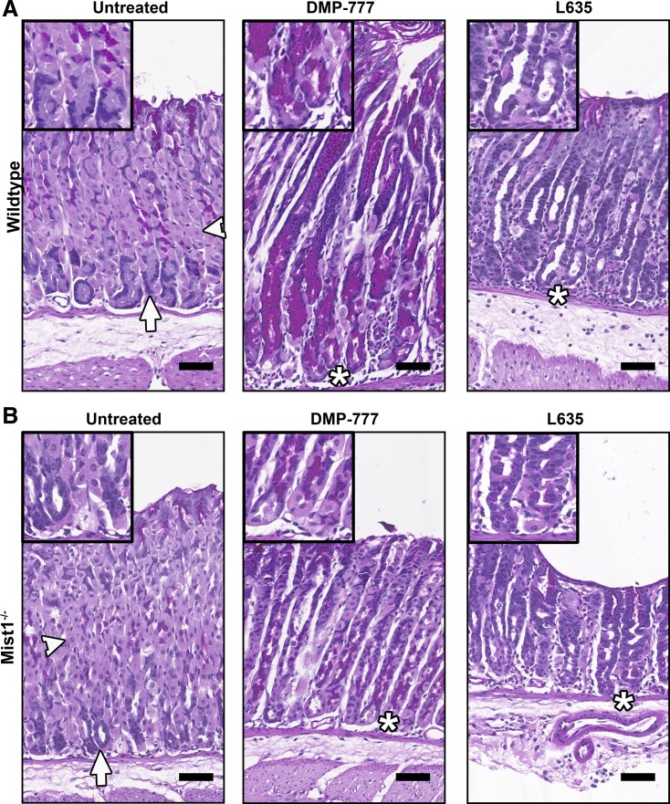
Fig. 1.
Periodic acid-Schiff (PAS) staining of untreated, DMP-777-treated, and L635-treated wild-type and Mist1−/− mice. DMP-777 or L635 was administered to wild-type mice (A) and Mist1−/− mice (B) for 14 or 3 days, respectively. DMP-777 and L635 treatment caused significant parietal cell loss (light pink, arrowheads) in both wild-type mice (A) and Mist1−/− mice (B). SPEM cells were identified as magenta-PAS-positive cells at the base of the glands (*), replacing the chief cells (blue, arrows) in drug-treated wild-type mice (A) and Mist1−/− mice (B). Note that L635-induced spasmolytic polypeptide-expressing metaplasia (SPEM) cells do not stain as bright magenta as DMP-777-induced SPEM cells. Scale bar = 50 µm.

