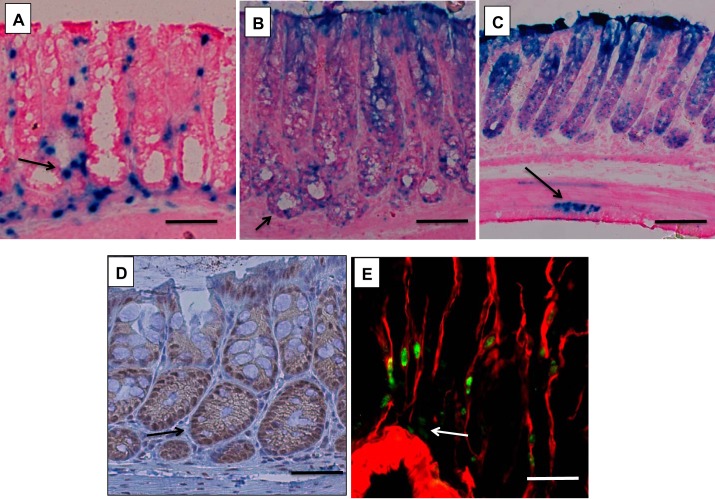
Fig. 1.
Expression of bone morphogenetic protein (BMP)-4 and localization of BMP signaling in the colon. A–C: X-Gal-stained frozen sections from BMP-4-β-galactosidase (β-gal)/+ mice (A) and BMP-responsive element (BRE)-β-gal mice (B and C). D: paraffin sections from wild-type mice stained with anti-p-Smad1, -5, and -8 antibodies and biotin-conjugated secondary antibodies. E: frozen sections from BMP-4-β-gal/+ mice stained with anti-β-gal primary antibodies and FITC-conjugated secondary antibodies (green), together with Cy3-conjugated-anti-actin, α-smooth muscle antibodies (red). Arrow in A points to BMP-4-expressing cells. Arrows in B, C, and D point to cells receiving BMP-generated signals. Arrow in E points to β-gal-positive α- smooth muscle actin (SMA)-negative cells. Results similar to those depicted were observed in at least three other separate experiments. Size bar is 50 μm in A, B, and D, 100 μm in C, and 20 μm in E.

