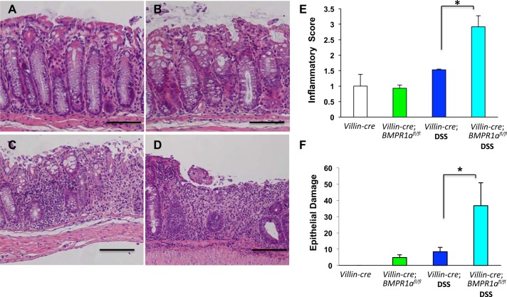
Fig. 5.
Loss of BMPR1A enhances inflammation and epithelial damage. A–D: representative H&E-stained gastric paraffin sections of the left colon of villin-Cre mice (A), villin-Cre;BMPR1Aflox/flox mice (B), DSS-treated villin-Cre mice (C), and of DSS-treated villin-Cre;BMPR1Aflox/flox mice (D). E and F: bar graphs represent inflammatory (E) and epithelial damage severity (F) scores calculated in both villin-Cre and villin-Cre;BMPR1Aflox/flox mice in the presence or absence of DSS. Values are shown as means ± SE, n = 3. *P < 0.05. Size bar = 100 μm.

