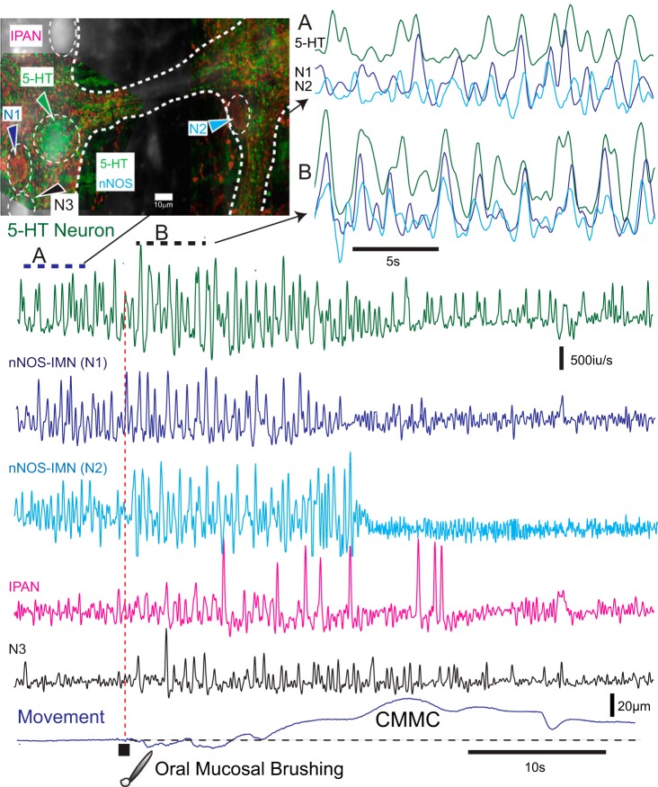
Fig. 3.
Activity in 5-hydroxytryptamine (5-HT) neurons and inhibitory motor neurons in Wnt1-GCaMP3 murine colon. In Wnt1-GCaMP3 mice, which labels all enteric neurons, subsequent 5-HT and neuronal nitric oxide synthase (nNOS) immunochemistry revealed a 5-HT neuron and two nNOS inhibitory motor neurons (IMNs, N1 and N2) in two different ganglia. These ganglia were in the midcolon. The myenteric neurons exhibited spontaneous Ca2+ transients that were differentiated to show Ca2+ spikes. Before the CMMC these neurons exhibited poorly coordinated activity (A, top right). Following brushing of the mucosa (five strokes with a brush in 1 s) at the oral end of the preparation, the 5-HT neuron and two nNOS-IMNs in the midcolon increased their activity, and their Ca2+ transients appeared more coordinated (B, bottom of top right) and coincided with the relaxation phase preceding the CMMC contraction. The nNOS-IMNs reduced their activity during the CMMC contraction. Following mucosal reflex stimulation, an intrinsic primary afferent neuron (IPAN; recognized by its ovoid shape and firing patterns) was transiently activated, as was another neuron (N3).

