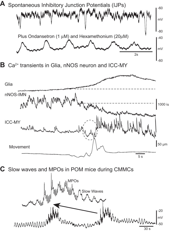
Fig. 4.
Spontaneous pacemaker activity and inhibitory junction potentials (IJPs). A: spontaneous fast IJPs in the circular muscle of murine colon recorded with intracellular microelectrodes. Following ondansetron (1 μM, a 5-HT3 receptor antagonist) and hexamethonium (20 μM, a nicotinic antagonist) administration tonic inhibition was abolished; that is, the membrane potential depolarized and spontaneous IJPs were abolished. These antagonists unmasked slow wave activity in the circular muscle. B: following a spontaneous contraction (bottom trace) Fluo-4 Ca2+ imaging revealed Ca2+ transients in a glial cell, adjacent IMNs, and neighboring myenteric pacemaker interstitial cells of Cajal (ICC-MY). The prolonged Ca2+ transient in the glial cell was associated with a reduction in spontaneous Ca2+ activity in an nNOS-IMN. During inhibition of the IMN, pacemaker myenteric potential oscillation (MPO) activity in ICC-MY turned on because it was under tonic inhibition (dotted circle indicates possible movement artifact). C: spontaneous CMMC-like activity in partially obstructed mice (POM) that lack tonic inhibition. Intracellular electrical recordings from circular muscle demonstrated that the circular muscle was depolarized and spontaneous IJPs were suppressed. Slow waves generated at the submucosal border by submucosal pacemaker interstitial cells of Cajal (ICC-SM) were observed to occur both between and during spontaneous CMMC-like activity. MPOs that originate in ICC-MY are readily observed during CMMC-like activity, where they summate with electrical slow waves originating in ICC-SM. However, CMMC-like contractions do not propagate in these mice, but occur synchronously along the colon, because they lack descending inhibition. (Dr. Dante Heredia performed the electrical recordings).

