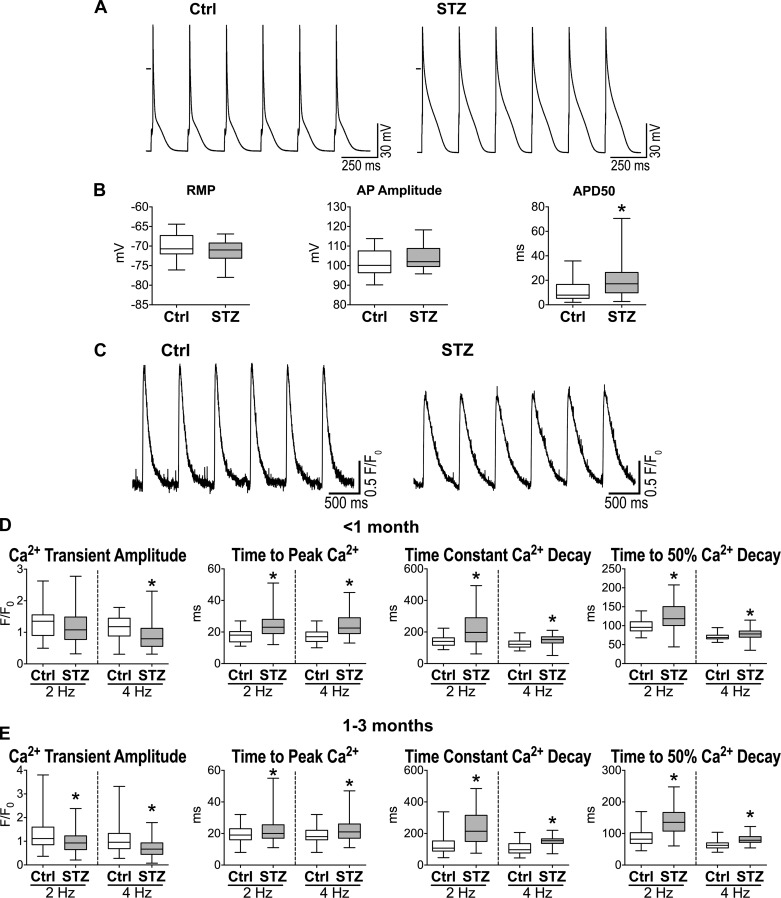
Fig. 5.
Diabetes induces defective myocyte function. A: action potentials (APs) of myocytes obtained from one naïve female C57Bl/6 mouse (Ctrl; left) and one STZ-treated female C57Bl/6 mouse 83 days after induction of diabetes (right). B: AP properties for isolated cardiomyocytes from naïve female C57Bl/6 mice (Ctrl, n = 27 cells from 13 animals) and STZ-treated female C57Bl/6 mice at 70–178 days after induction of diabetes (n = 28 cells from 8 animals) are shown as median and interquartile ranges. *P < 0.05 vs. Ctrl. RMP, resting membrane potential; APD50, AP duration at 50% repolarization. C: Ca2+ transients in myocytes obtained from one naïve female C57Bl/6 mouse (Ctrl; left) and one STZ-treated female C57Bl/6 mouse 60 days after induction of diabetes (right). D: Ca2+ transient properties obtained at 2- and 4-Hz pacing rates for isolated cardiomyocytes from naïve female C57Bl/6 mice (Ctrl, n = 35–38 cells from 2 animals) and STZ-treated female C57Bl/6 mice at 15–23 days after induction of diabetes (n = 64–71 cells from 4 animals) are shown as median and interquartile ranges. *P < 0.01 vs. Ctrl. E: Ca2+ transient properties obtained at 2- and 4-Hz pacing rate for isolated cardiomyocytes from naïve female C57Bl/6 mice (Ctrl, n = 153 cells from 9 animals) and STZ-treated female C57Bl/6 mice at 38–82 days after induction of diabetes (n = 188–189 cells from 9 animals) are shown as median and interquartile ranges. *P < 0.001 vs. Ctrl.

