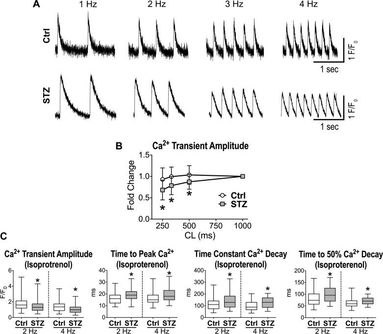
Fig. 6.
Diabetes affects rate adaptation and inotropic mechanisms of cardiomyocytes. A: Ca2+ transients at progressively faster pacing rate for one myocyte obtained from a naïve C57Bl/6 female mouse (Ctrl; top) and one myocyte from a STZ-treated female C57Bl/6 mouse 38 days after induction of diabetes (bottom). B: quantitative data for rate adaptation of amplitude and decay of Ca2+ transients for cardiomyocytes from naïve female C57Bl/6 mice (Ctrl, n = 24 cells from 5 animals) and STZ-treated female C57Bl/6 mice at 38–88 days after induction of diabetes (STZ, n = 16 cells from 4 animals) are shown as means ± SE. *P < 0.05 vs. Ctrl at the same cycle length (CL). C: Ca2+ transient properties obtained at 2- and 4-Hz pacing rates in the presence of 100 nM isoproterenol for myocytes from naïve female C57Bl/6 mice (Ctrl, n = 88 cells from 5 animals) and STZ-treated female C57Bl/6 mice at 40–82 days after induction of diabetes (n = 85–86 cells from 5 animals) are shown as median and interquartile ranges. F/F0, normalized fluorescence. *P < 0.01 vs. Ctrl.

