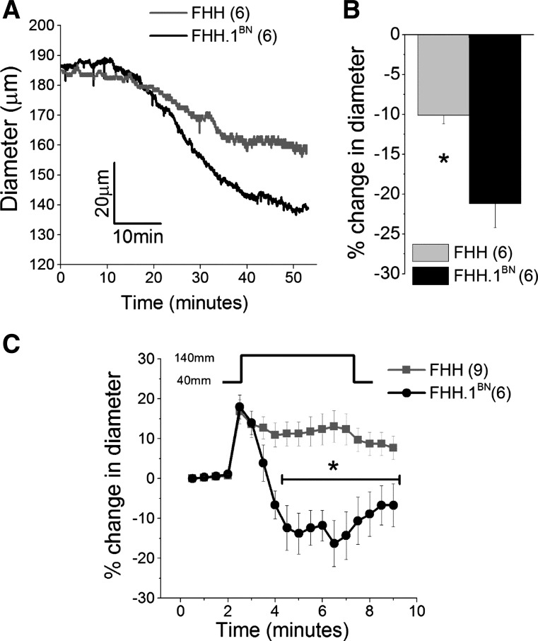
Fig. 1.
Comparison of basal tone and spontaneous myogenic response in middle cerebral arteries (MCAs) isolated from FHH and FHH.1BN rats. MCAs were mounted on glass micropipettes in a myograph, and intraluminal pressure was maintained at 80 mmHg. A and B: traces and a comparison of the change in the diameter of the MCAs during equilibration period at 37°C. C: percent change in diameter of MCAs plotted over time in response to a step change in luminal pressure from 40 to 140 mmHg spontaneously. *Significant difference in the corresponding values in FHH and FHH.1BN rats. Error bars ± SE. Numbers in parentheses indicate the number of vessels studied.

