Fig. 6.
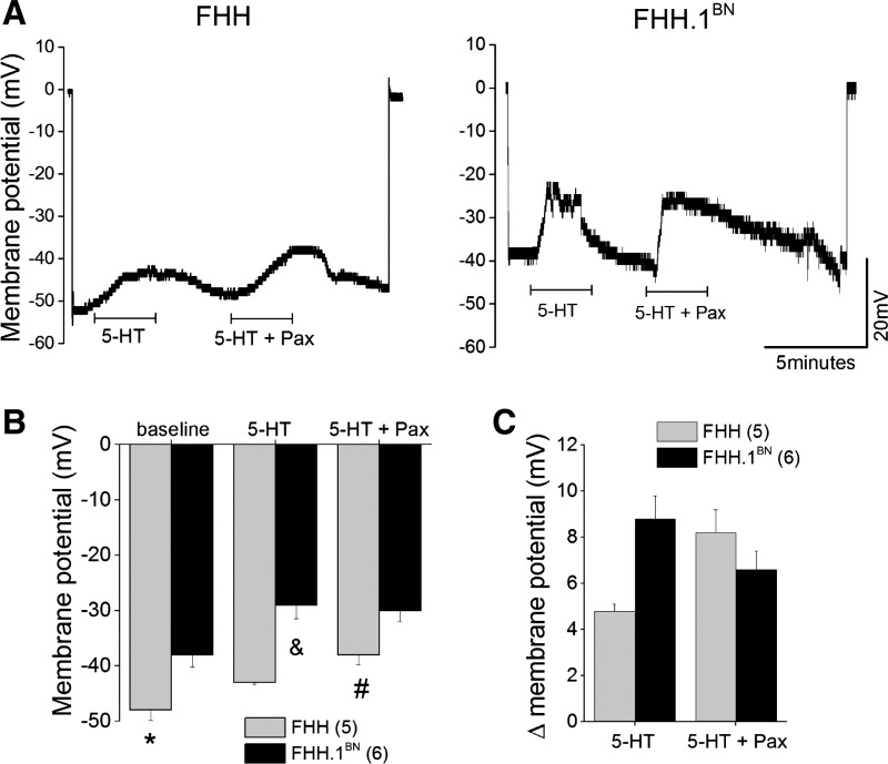
Comparison of 5-HT-mediated change in membrane potential in pressurized MCAs isolated from FHH and FHH.1BN rats. Membrane potential was measured in pressurized (80 mmHg) MCAs using microelectrode impalement technique. A and B: traces of the original membrane potential recordings and summary bar graph of the membrane potential before and after application of either 5-HT (1 μM) alone or combined with paxilline (100 nM). C: delta change in the membrane potential before and after application of 5-HT alone or combined with paxilline. Values are means ± SE. *Significant difference in the corresponding values in FHH and FHH.1BN rats; &significant difference in the corresponding values from the 5-HT-mediated depolarization in FHH.1BN rats; #significant difference in the corresponding values from the 5-HT + paxilline-mediated depolarization in FHH rats. Numbers in parentheses indicate the number of vessels studied.

