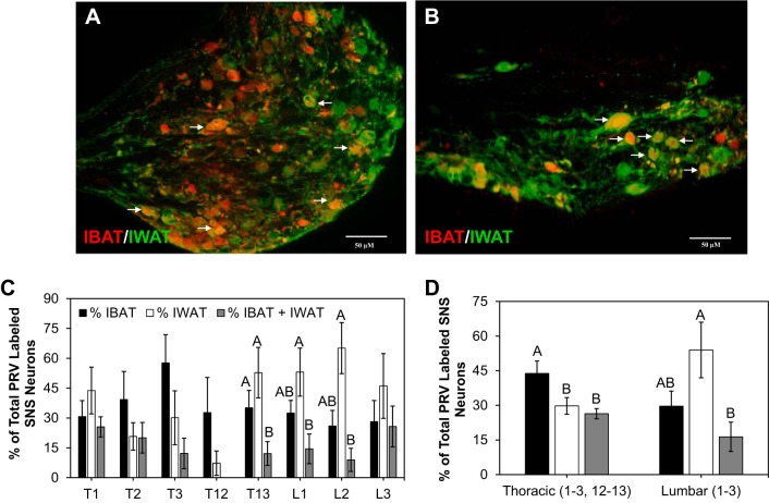
Fig. 1.
Distribution of sympathetic nervous system (SNS) neurons to interscapular brown adipose tissure (IBAT) and inquinal white adipose tissue (IWAT) across thoracic and lumbar regions of the SNS ganglia. A and B: representative pseudorabies virus (PRV) labeling of SNS neurons to IBAT (red) and IWAT (green) in T3 (A) and L3 (B) SNS ganglia. Doubly labeled neurons are indicated by white arrows. C: percentage of total PRV-labeled SNS neurons across each thoracic (T1–3, 12–13) and lumbar (L1–3) regions. D: percentage of total PRV-labeled SNS neurons across collapsed thoracic and lumbar regions. Scale bar = 50 µm. Values that do not share a common superscript are significantly different at P < 0.05.

