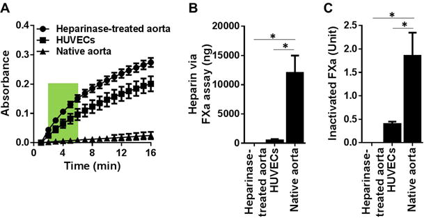
Figure 3. Measurement of 1 cm2 biological surface’s capacity to form pro-coagulant complexes and heparin equivalent estimation with the FXa assay.

(A) Absorbance readings of residual FXa generated from: native aorta, heparinase-treated aorta and HUVEC monolayer of 1 cm2 surface area. The results are mean ± SEM of 5 independent assays using five different rats and HUVEC culture (n=5). (B) Heparin equivalent weight was estimated via the slope of curve at 2–6 min (green box in A) against the standard curve in Figure 1B. (C) Inactivated FXa was estimated via the absorbance at 16 min (end point in A) against the standard curve in Figure 1C. * p < 0.05.
