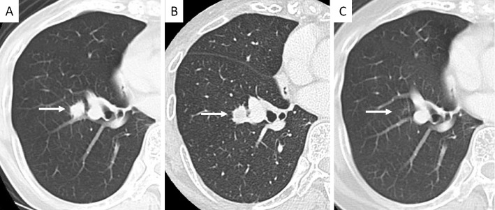
Figure 2.
A: Chest computed tomography showed a solitary nodule (12 mm in diameter) in the right lower lobe just before undergoing biopsy (arrow). B: Three months after the biopsy, the diameter of the nodule has slightly increased (arrow). C: Fifteen months after the biopsy, the nodule has disappeared (arrow).

