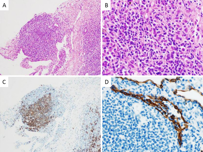
Figure 3.
A: Histological examinations showed an accumulation of small lymphoid cells [Hematoxylin and Eosin (H&E) staining, ×40]. B: Lymphoid cells consist of atypical small lymphocytes (H&E staining, ×200). C: Cells in the lymphatic follicle include clusters of differentiation (CD) 20 positive (CD20 stain, ×40). D: Infiltration of lymphoid cells into the epithelium, defined as lympho-epithelial lesion (LEL) (Cytokeratin stain, ×200).

