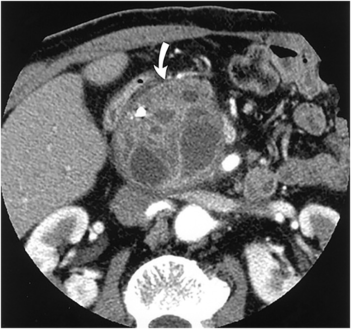
Figure 4.
Contrast-enhanced CT scan showing branch duct IPMN. A cystic lesion can be seen with a markedly dilated side branch duct with wall thickening (arrow). (Adapted with permission from: Kim YH et al.)68 IPMN, intraductal papillary mucinous neoplasm.

